Solving the Structural Variation Analysis Roadblock
Structural variants (SVs) are behind the diseases we study most closely in genomics research—cancer, neurological and neurodegenerative disease, cardiovascular conditions, inherited disorders—through large deletions, duplications, inversions, and rearrangements that reshape genome architecture. Despite that reach, SVs remain one of the least understood categories of genomic variation. A substantial share of rare disease cases still have no identified genetic cause after sequencing, with estimates ranging from roughly 25–80% depending on cohort and method, and SVs are a recognized piece of that gap.
It's not for lack of effort. Short-read sequencing has had two decades to mature, and it has become remarkably good at calling single-nucleotide variants. SNV detection and SV detection, though, are fundamentally different problems, and a tool optimized for one isn't necessarily well suited to the other.
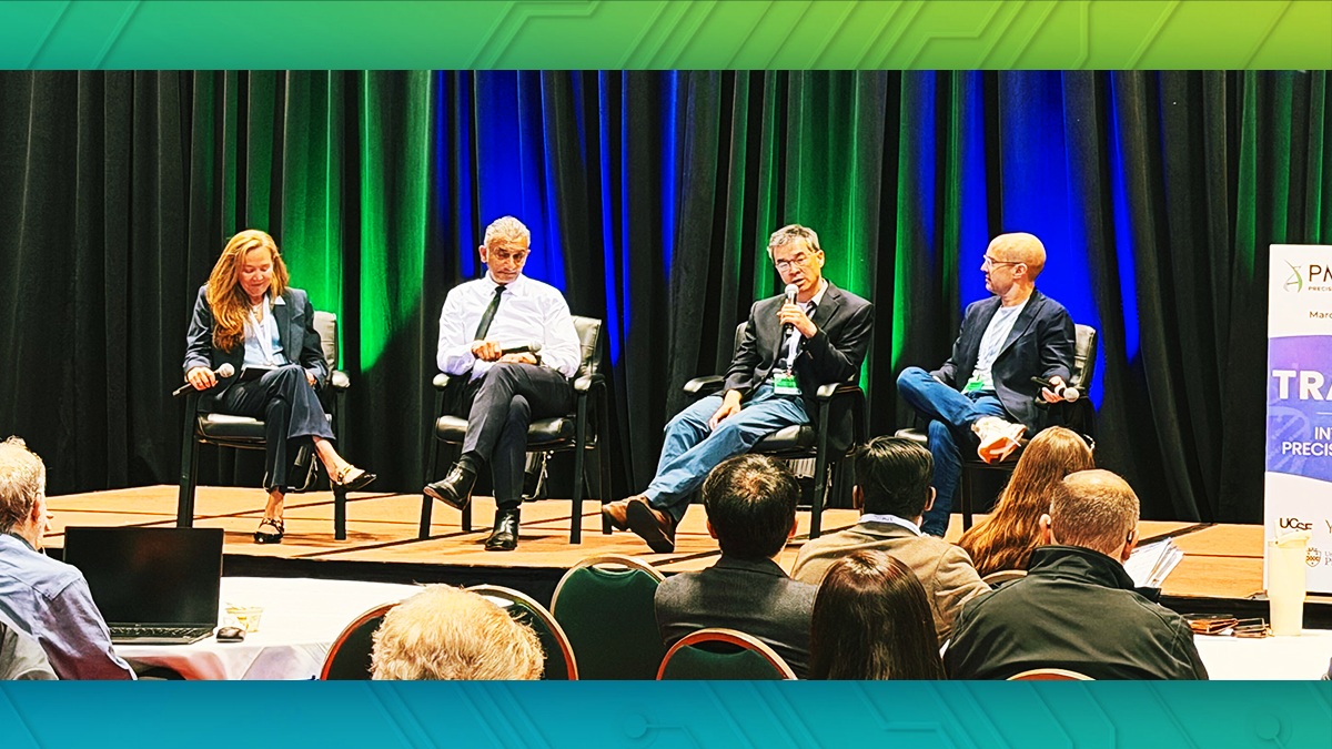
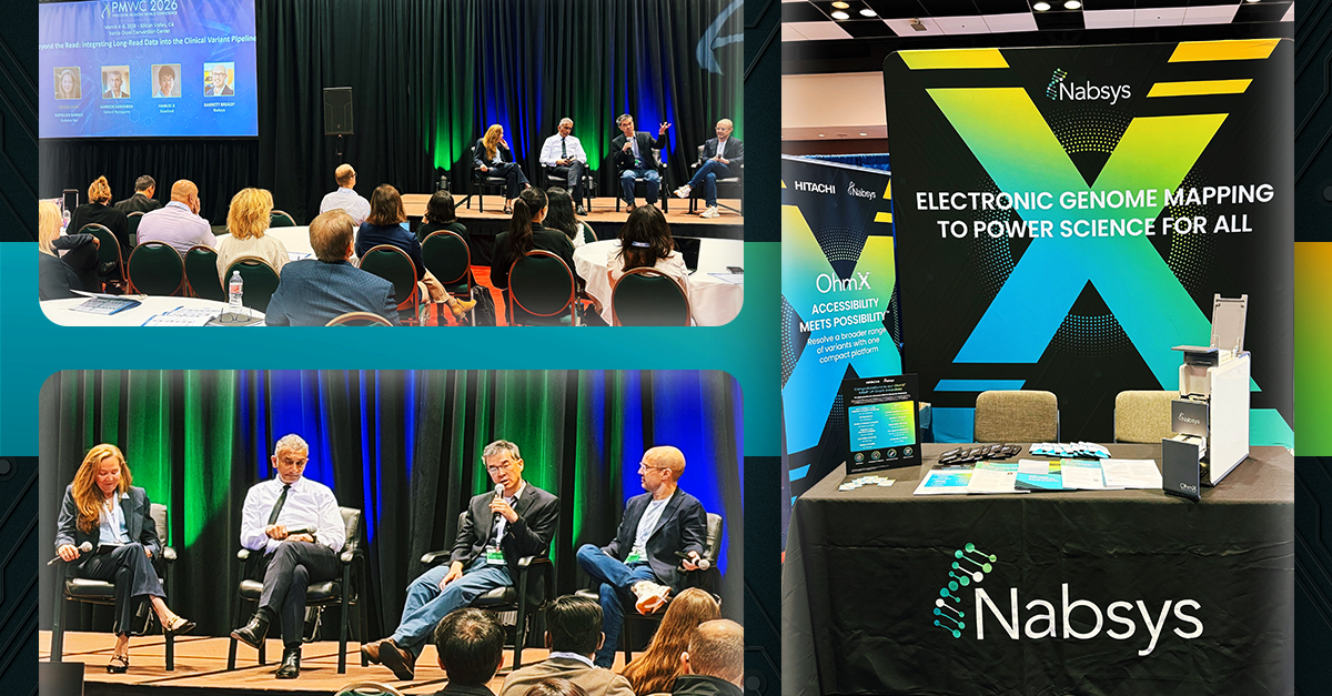
That gap was the subject of a panel discussion at the Precision Medicine World Conference (PMWC) in Santa Clara. There, Nabsys founder and CEO Barrett Bready, M.D., joined Gordon Sanghera, Ph.D., co-founder and CEO of Oxford Nanopore, and Hanlee Ji, M.D., Professor of Medicine at Stanford, to talk through where SV detection stands today and what it will take to translate that research toward broader impact.
PMWC brings together genomics researchers, technology developers, and translational scientists to take stock of where the field is moving. The 2026 panel drew perspectives from oncology, sequencing technology, and genome mapping to approach the problem from multiple angles.
Why the Standard Toolkit Falls Short
A short read runs 150 to 300 base pairs. SVs run from 50 base pairs to millions. A fragment that short struggles to span a variant that large. SVs also tend to sit in repetitive stretches of the genome, so short-read alignment ends up ambiguous in exactly the regions where precision matters most. Large insertions can be missed entirely. Complex rearrangements get inferred from surrounding evidence instead of being directly resolved.
Karyotyping has covered this territory for decades, and it's still widely used today, but it was designed for a visual snapshot, not molecular resolution, and it's slow. Long-read sequencing, microarrays, and genome mapping have each stepped in to modernize it. Dr. Bready doesn't see this as a contest between them:
"The challenge isn't just to detect SVs, but to also understand what additional information we need to make sense of them. With multiple technologies converging on the SV analysis problem, there will always be trade-offs. Our goal is to create next-generation cytogenetics—a modern evolution of traditional chromosome analysis with higher resolution and accessibility. The point isn't which technology wins, but how they can be integrated to complement each other."
Why This Matters Now
This isn’t just academic interest. SVs are turning up as some of the field’s most actionable drug targets and markers of disease biology. Dr. Ji, who studies SV patterns in cancer at Stanford, described what improved resolution makes accessible from a research standpoint:
"With these types of structural variant and long-range genomic analyses, we can probably tease out all types of rearrangements that are just completely unknown. So as a drug target, that's a natural go-to."
Better detection alone won't move SV analysis into standard research practice. It also takes validation and a shared framework for interpreting SVs—the kind ACMG and AMP standards already provide for sequence variants. Dr. Ji again:
"There's already recognition in the general community that these types of large structural events are important."
Gordon Sanghera, Ph.D., co-founder and CEO of Oxford Nanopore, made a related point about how much is still unaccounted for. By his account, structural variation, copy number variation, and methylation together explain more than a quarter of rare diseases that remain genetically unresolved, helping explain why research on conditions like neurodegeneration has advanced slowly. On the pace the field needs to pick up, he didn't mince words: "we already have 25% of diseases unmapped with short read," and closing that gap "will radically transform drug discovery."
Rethinking the Cost Equation
Long-read, single-molecule sequencing earns its power the hard way: one molecule at a time. Short-read sequencing works differently, sequencing huge numbers of fragments in parallel as an ensemble. Dr. Bready put it down to a basic physics difference. Short-read sequencing works as an ensemble, while long-read sequencing must be single-molecule. He called it, plainly, "a harder physics problem to solve."
Given that, the fix probably isn't one platform trying to do it all. Run low-cost short-read sequencing for SNVs and small variants, and pair it with a low-cost, high-resolution SV tool for the rest. Together they cover the genome without single-molecule pricing across the board. That's the gap electronic genome mapping (EGM) is built to fill—bringing SV resolution down to a price where it fits into routine workflows instead of getting reserved for the hardest cases.
Where Electronic Genome Mapping Comes In
Nabsys built the OhmX™ Platform, powered by EGM, for exactly this problem.
In EGM, ultra-long DNA molecules travel through solid-state nanochannels lined with electronic sensors. Site-specific tags along the molecule generate voltage changes as they pass through, and those electronic signals get assembled into dense, long-range genome maps.
The electronic method changes more than the mechanism—it changes the economics. Optical mapping depends on imaging DNA and reading fluorescent signals, which ties it to the resolution limits of light diffraction and the cost of the imaging hardware needed to support it. As Dr. Bready described it, optical technologies "depend on the optical properties of the analyte." Electronic detection doesn't carry that constraint. It "depends only on the shape of the analyte" moving through the channel. That's why EGM's solid-state detector is stable, reusable, and runs at room temperature, with no laser or imaging system to maintain. It also lets the OhmX Platform run in a smaller footprint than optical genome mapping instruments, at 2–5× lower cost.
EGM was designed to enable genome-wide, de novo SV discovery, using the same electronic detection process described above. Today, one of its most established uses is as an orthogonal layer of confirmation alongside NGS, long-read sequencing, karyotyping, and microarray data. On that application, Dr. Bready said:
"The ability for EGM to provide orthogonal validation or confirmation of SV detection is incredibly important. When you have the ability to confirm some of these structural variants—many of which were undetected with legacy technologies—it makes everybody more comfortable using the data going forward."
The longer-term aim is broader: running EGM alongside NGS not just to confirm what other platforms find, but as a routine discovery pairing in its own right, contributing new SV calls as a standard part of the workflow rather than a secondary check.
The Path to Next-Generation Cytogenetics
Nabsys built EGM on a straightforward idea: structural variation needed a technology designed for it, rather than an adaptation of tools built for a different question. As orthogonal confirmation becomes routine and interpretation standards catch up, the question for labs stops being whether to add SV resolution and starts being how fast they can get it running.
That's what EGM and the OhmX Platform are here for.
When Sanghera called it “a golden age of multiomics,” he wasn’t describing a distant horizon. The tools, the standards, and the community momentum are converging now. Researchers who build multi-technology SV workflows today will be the ones making the discoveries that reshape what we know about disease biology.
Watch the full PMWC panel recording.